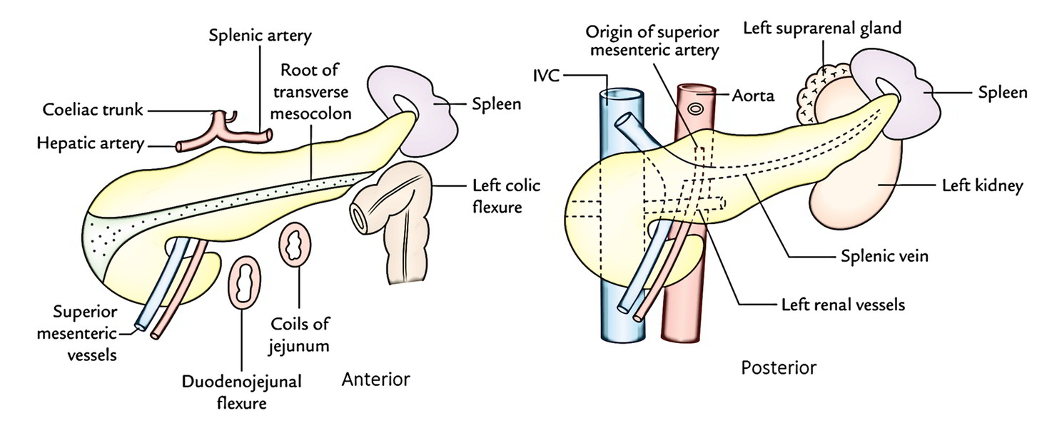
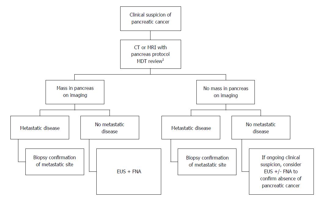
After taking a medical history and performing a physical exam your doctor may recommend imaging tests to help with diagnosis and treatment planning. The upper limits of a normal duct diameter on ct is considered to be 2 to 3 mm in the body and tail and up to 5 mm in the head.
Imaging Preoperatively For Pancreatic Adenocarcinoma
Pancreatic body measurements. The diameter of the main pancreatic duct is a commonly assessed parameter in imaging. On ercp the duct is 3 to 4 mm in the head 2 to 3 mm in the body and 1 to 2 mm in the tail. The duct diameter is greatest at the head and neck region and is slightly narrower towards the body and tail. The diameter of duct can increase with inspiration 3. The average maximum diameter of the main pancreatic duct autopsy series was 29 mm 50yrs and 35 mm 50years. In mini pigs the measurements of pancreatic volume by mri and by water displacement were almost identical r2 09867.
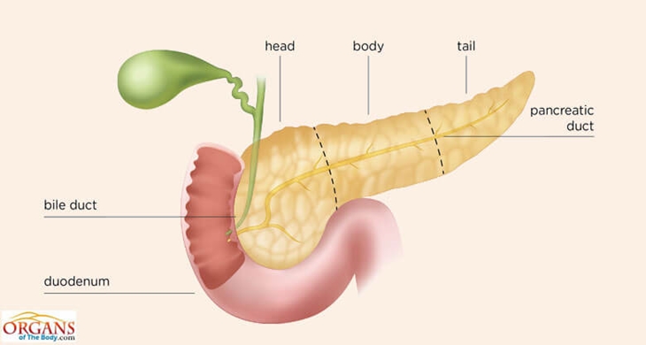
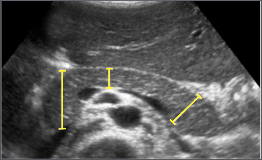
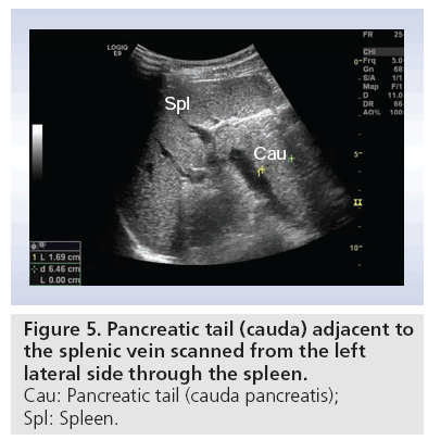
The initial fields included a margin of more than 5 cm of uninvolved esophagus superiorly a margin of about 5 cm of stomach in all directions and nodal groups including celiac artery and lesser curvature nodes suprapancreatic bodytail of pancreas and splenic hilum. The size of the normal pancreas was found to be up to 30 cm for the head 25 cm for the neck and body and 20 cm for the tail. Approximate normal measurements are. The head of the pancreas is on the right side of the abdomen and is connected to the duodenum the. Pancreatic size in each anatomical section was smaller when measured by us than by mri table 2. 12 20 cm pancreatic duct.
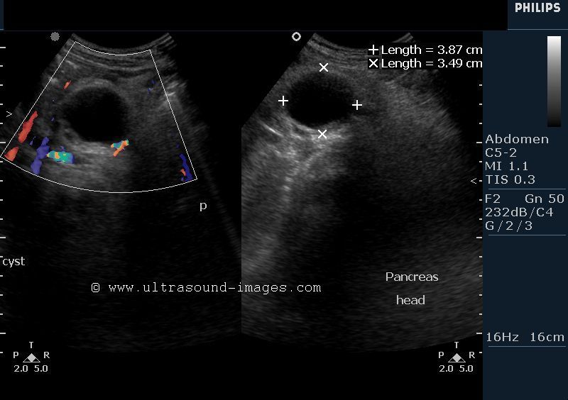
In humans the average pancreas volume was 72745 ml range from 350 to 1055 ml. Its normal reported value ranges between 1 35 mm 58. The mean difference between mri and us measurements was 039 cm for the head 018 cm for the body and 054 cm for the tail which corresponds to a relative difference of 144433 figure 1. This result is in strong agreement with results of previous large postmortem and computed tomography ct studies. Head 35mm anterior to posterior neck 10 15mm tail 20mm. Pancreatic cysts are diagnosed more often than in the past because improved imaging technology finds them more readily.
The pancreas is about 6 inches long and sits across the back of the abdomen behind the stomach. Many pancreatic cysts are found during abdominal scans for other problems.
